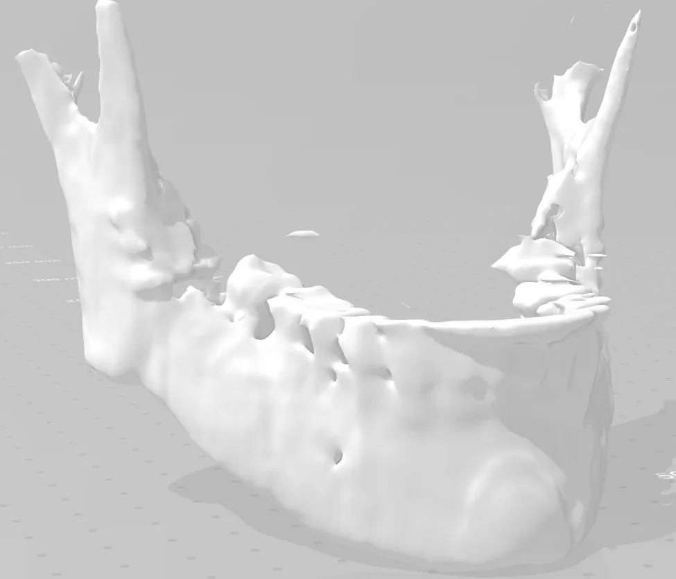
Through the use of a tomography, the aim is to create a three-dimensional model of a jaw, in order to help dentists to be able to perform dental surgeries before carrying them out on real patients, in addition to the creation and analysis of jaw prostheses.
Understanding the Reconstruction
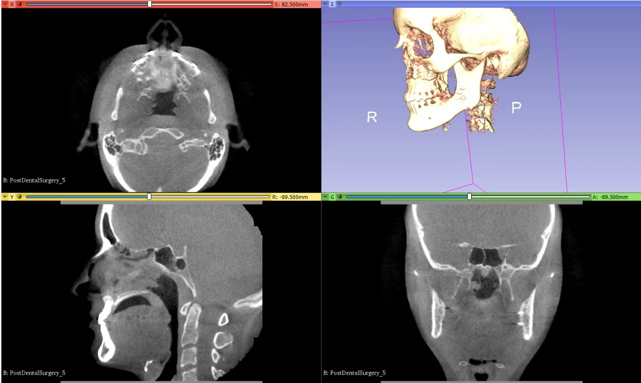
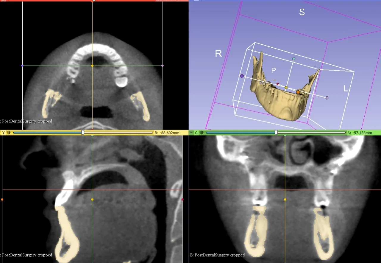
The workflow converts volumetric images into a segmented surface for printing. Spacing, segmentation, smoothing and export units affect geometry. The screenshots document a modelling project; the clinical and prosthetic applications below are motivations, not evidence of clinical validation or a patient-ready implant.
Testing and Real-World Use
This could be tested with a synthetic volume by checking voxel spacing and comparing known dimensions before surface export and after printing. Geometric traceability is useful for anatomical-model research, but this project does not establish suitability for diagnosis, surgery or implant design.
Objective
• Printing of the 3D model of the jaw for practice and help in operating techniques. • Improvements in prototyping material and application to prostheses.
Methodology
graph TB;
    A001[Computed tomography of patients.]
    A002[Image processing for 3D model creation.]
    A003[Noise removal in the three-dimensional model.]
    A004[Conversion to STL format of the model.]
    A005[Mechanical simulations on the model.]
    A006[3D model printing.]
    A007[Mechanical analysis of the model.]
    A008[Use of model by dentists.]
    A009[Improvements in the manufacture of jaws and plates for prostheses of these.]
    A001-->A002;
    A002-->A003;
    A003-->A004;
    A004-->A005;
    A005-->A006;
    A006-->A007;
    A007-->A008;
    A008-->A009;
Results
STL